User danghoainhan012 uploaded the image
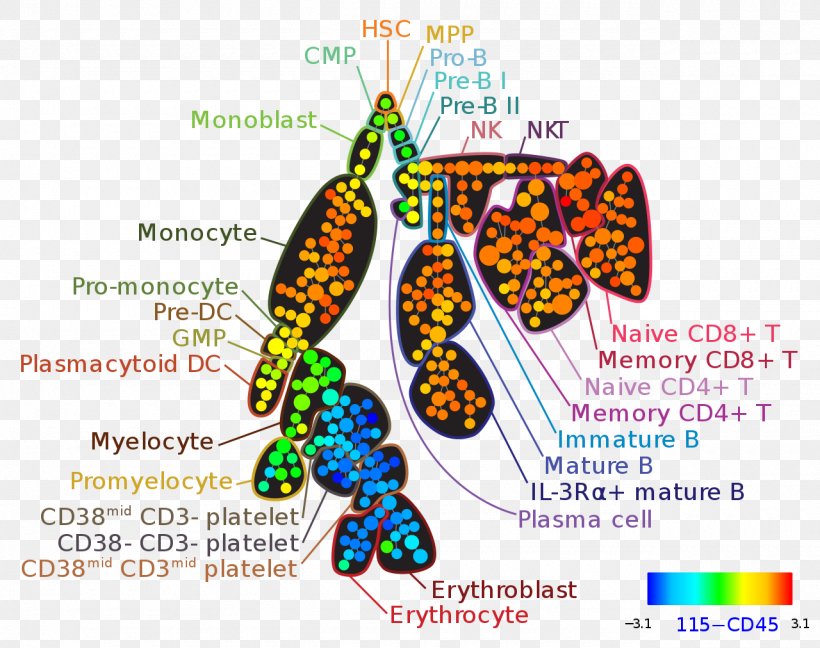
A diagram that shows the structure of a protein molecule. It is divided into different sections, each representing a different type of protein. The first section is labeled "Monoblast" and shows the different types of proteins that make up the protein molecule, such as CMP, HSC, MPP, Pro-B, and NK. The proteins are arranged in a circular pattern, with each protein represented by a different color. The colors range from orange, yellow, green, blue, and purple, representing the different levels of protein in the molecule. In the center of the image, there is a large orange and yellow molecule, which appears to be the nucleus of the protein. The nucleus is surrounded by smaller orange and green molecules, which are connected by a series of interconnected lines. The molecules are labeled with their names, including "CMP", "Pro-B", "NK", and "NK". On the left side of the diagram, there are several smaller orange molecules, including the nucleus, mitochondria, and mitochondria. On the right side, the nucleus is labeled as "Naive CD8+ T", which are the nuclei of the nucleus. The nuclei are labeled as the nucleis of the mitochondria and the mitochondrion. The mitochondria is labeled with the names of the different nuclei, including DNA, RNA, and DNA. - The diagram also shows the number of nuclei in each molecule, as well as the number in each cell. The image also includes a color-coded bar at the bottom right corner, which shows that the molecule is 115-CD45.
Mass Cytometry Flow Cytometry Bioinformatics High Throughput Biology PNG
. The resolution of this PNG file is 1280 x 1012 pixels and it has a file size of 350.67 KB.A diagram that shows the structure of a protein molecule. It is divided into different sections, each representing a different type of protein. The first section is labeled "Monoblast" and shows the different types of proteins that make up the protein molecule, such as CMP, HSC, MPP, Pro-B, and NK. The proteins are arranged in a circular pattern, with each protein represented by a different color. The colors range from orange, yellow, green, blue, and purple, representing the different levels of protein in the molecule. In the center of the image, there is a large orange and yellow molecule, which appears to be the nucleus of the protein. The nucleus is surrounded by smaller orange and green molecules, which are connected by a series of interconnected lines. The molecules are labeled with their names, including "CMP", "Pro-B", "NK", and "NK". On the left side of the diagram, there are several smaller orange molecules, including the nucleus, mitochondria, and mitochondria. On the right side, the nucleus is labeled as "Naive CD8+ T", which are the nuclei of the nucleus. The nuclei are labeled as the nucleis of the mitochondria and the mitochondrion. The mitochondria is labeled with the names of the different nuclei, including DNA, RNA, and DNA. - The diagram also shows the number of nuclei in each molecule, as well as the number in each cell. The image also includes a color-coded bar at the bottom right corner, which shows that the molecule is 115-CD45.
Mass Cytometry Flow Cytometry Bioinformatics High Throughput Biology PNG
Related PNG Images