User palanthas uploaded the image
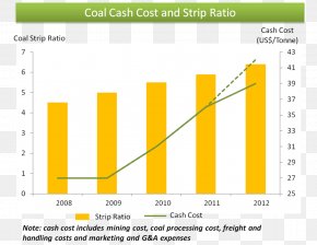
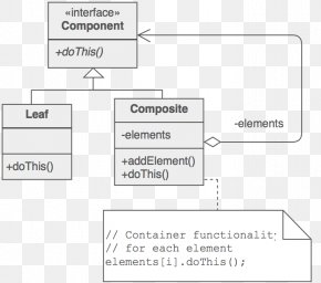
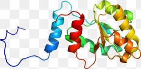
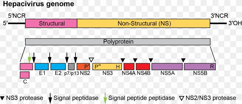
A diagram that shows the structure of a Hepacivirus genome. It shows the different types of proteins that are responsible for the production of Hepac virus genes. The diagram is divided into three sections. The top section is labeled "Structural" and shows the number of proteins in each section. The structure is 5NCR, which is a type of protein that binds to the structure. The second section is titled "Non-Structural (NS)". The third section is labelled "Polyprotein" and has a label that reads "3NCR". Below the structure, there is a bar graph that shows that the protein is composed of different colors - pink, blue, green, yellow, and red. The colors are arranged in a horizontal line, with the pink color representing the structure and the blue color representing a protein. The red color represents the protein, while the yellow color represents a protein with a protein in the center. The graph also shows that there are three proteins in the structure - E1, E2, E3, E4, E5, E6, E7, E8, E9, E10, E11, E12, E13, E14, E15, E16, E17, E18, E19, E20, E21, E22, E23, E24, E25, E26, E27, E28, E29, E30, E31, E32, E33, E34, E35, E36, E37, E38, E39, E40, E41, E42, E43, E44, E45, E46, E47, E48, E50, E51, E52, E53, E54, E55, E56, E57, E58, E60, E61, E62, E63, E64, E65, E66, E67, E68, E69, E70, E71, E72, E73, E74, E75, E76, E77, E78, E79, E80, E81, E82, E83, E84, E85, E86, E87, E88, E90, E91, E92, E93, E94, E95, E96, E97, E98, E99, E.
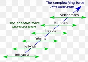
Angle Document Line Purple Special Olympics Area M PNG
. The resolution of this PNG file is 1660 x 718 pixels and it has a file size of 42.65 KB.A diagram that shows the structure of a Hepacivirus genome. It shows the different types of proteins that are responsible for the production of Hepac virus genes. The diagram is divided into three sections. The top section is labeled "Structural" and shows the number of proteins in each section. The structure is 5NCR, which is a type of protein that binds to the structure. The second section is titled "Non-Structural (NS)". The third section is labelled "Polyprotein" and has a label that reads "3NCR". Below the structure, there is a bar graph that shows that the protein is composed of different colors - pink, blue, green, yellow, and red. The colors are arranged in a horizontal line, with the pink color representing the structure and the blue color representing a protein. The red color represents the protein, while the yellow color represents a protein with a protein in the center. The graph also shows that there are three proteins in the structure - E1, E2, E3, E4, E5, E6, E7, E8, E9, E10, E11, E12, E13, E14, E15, E16, E17, E18, E19, E20, E21, E22, E23, E24, E25, E26, E27, E28, E29, E30, E31, E32, E33, E34, E35, E36, E37, E38, E39, E40, E41, E42, E43, E44, E45, E46, E47, E48, E50, E51, E52, E53, E54, E55, E56, E57, E58, E60, E61, E62, E63, E64, E65, E66, E67, E68, E69, E70, E71, E72, E73, E74, E75, E76, E77, E78, E79, E80, E81, E82, E83, E84, E85, E86, E87, E88, E90, E91, E92, E93, E94, E95, E96, E97, E98, E99, E.
Related PNG Images